diabetic foot maggots
The feet and hands are most commonly affected. Maggots are placed on the wound followed by wrapping with secondary dressing.

Use Of Lucilia Sericata Blowfly Maggots In The Treatment Of Diabetic Feet Threatened With Amputation Semantic Scholar
Where is it located.
. In 2004 the United States Food and Drug Administration. In diabetic foot ulcers there is tentative evidence of benefit. Sterile maggots of the green bottle fly Lucilia sericata are placed directly into the affected area and held in place by a close net dressing.
Diabetic neuropathy vascular structures superficial deep phlebitis pitting oedema lipodermatosclerosis and contact dermatitis. Symptoms may include a change in skin color to red or black numbness swelling pain skin breakdown and coolness. Hyperbaric oxygen therapy has proved effective in treating gangrene caused by infected diabetic foot ulcers reducing the risk of amputation.
Venous hypertension as a result of venous reflux incompetence or. Because maggots will only eat dead tissue they can be applied to the affected tissues and allowed to eat away the dead skin. Try to keep an open mind about maggot therapy.
Measurements-in centimeters Length X Width X Depth Length greatest length head to toe. Wounds that have necrotic tissue present will not heal therefore one of the above methods will be required to remove the devitalized tissue. If the gangrenous area is too large for debridement a surgeon may need to amputate the affected part such as the foot or fingers.
Thank you for making Chowhound a vibrant and passionate community of food trailblazers for 25 years. Maggot therapy has been effective in the treatment of diabetic foot ulcers. A Cochrane review of methods for the debridement of venous leg ulcers found maggot therapy to be broadly as effective as most other methods but the study also noted that the quality of data was poor.
The maggots are placed on the wound and covered with gauze under a firm dressing which keeps them on the wound and out of sight. Venous ulcers are the most common type of chronic lower extremity ulcers affecting 1 to 3 of the US. Biosurgical debridement using larval therapy maggots.
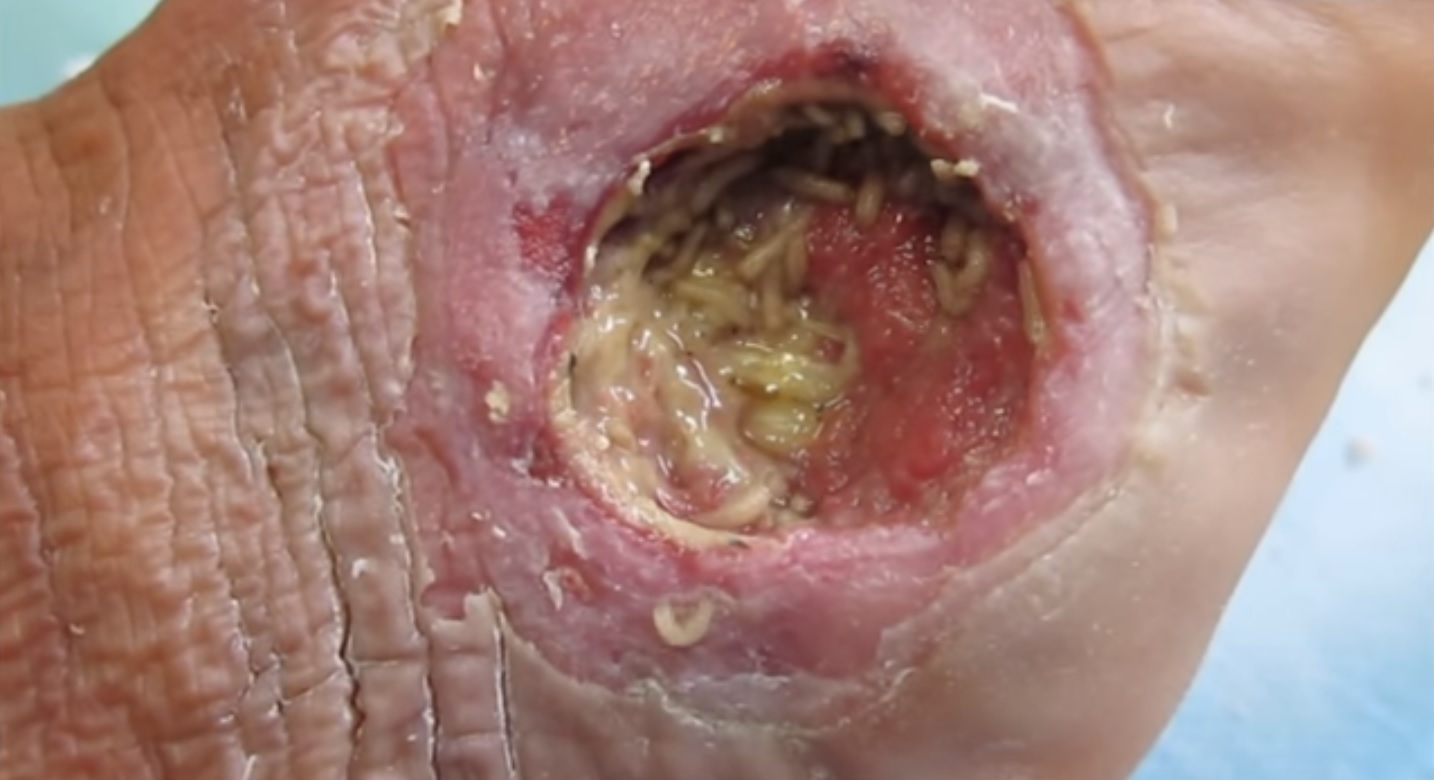
Necrosis can be caused by a number of external sources including injury infection cancer infarction poisons and inflammation. Surgical debridement of a diabetic foot ulcer stimulates the edge of the wound releases growth factors and reduces inflammation. The larvae feed on the necrotic dead tissue and bacteria present at the wound site and secrete antimicrobial enzymes.
If the gangrene is caused by an infectious agent it may present with a fever or sepsis. If gangrene occurs because of poor blood circulation a surgeon may perform vascular surgery to the blood vessels to try to improve the circulation and keep the tissue as healthy as possible. After a few days the dressing is cut away and the maggots are removed.
Diabetic foot ulcer DFU is the most costly and devastating complication of diabetes mellitus which affect 15 of diabetic patients during their lifetime. Sterile lab-raised maggots are often used to treat gangrene. Maggot Therapy - A small number of a special species of maggots are introduced into the ulcer.
These eat only the dead skin and produce chemicals that promote healing. This process can also help your body to heal itself and help to prevent infection. Many patients find the mere idea of maggot therapy distasteful- obviously these patients are not suitable candidates for this type of therapy.
Maggot therapy improves healing in chronic ulcers. We wish you all the best on your future culinary endeavors. Gangrene is a type of tissue death caused by a lack of blood supply.
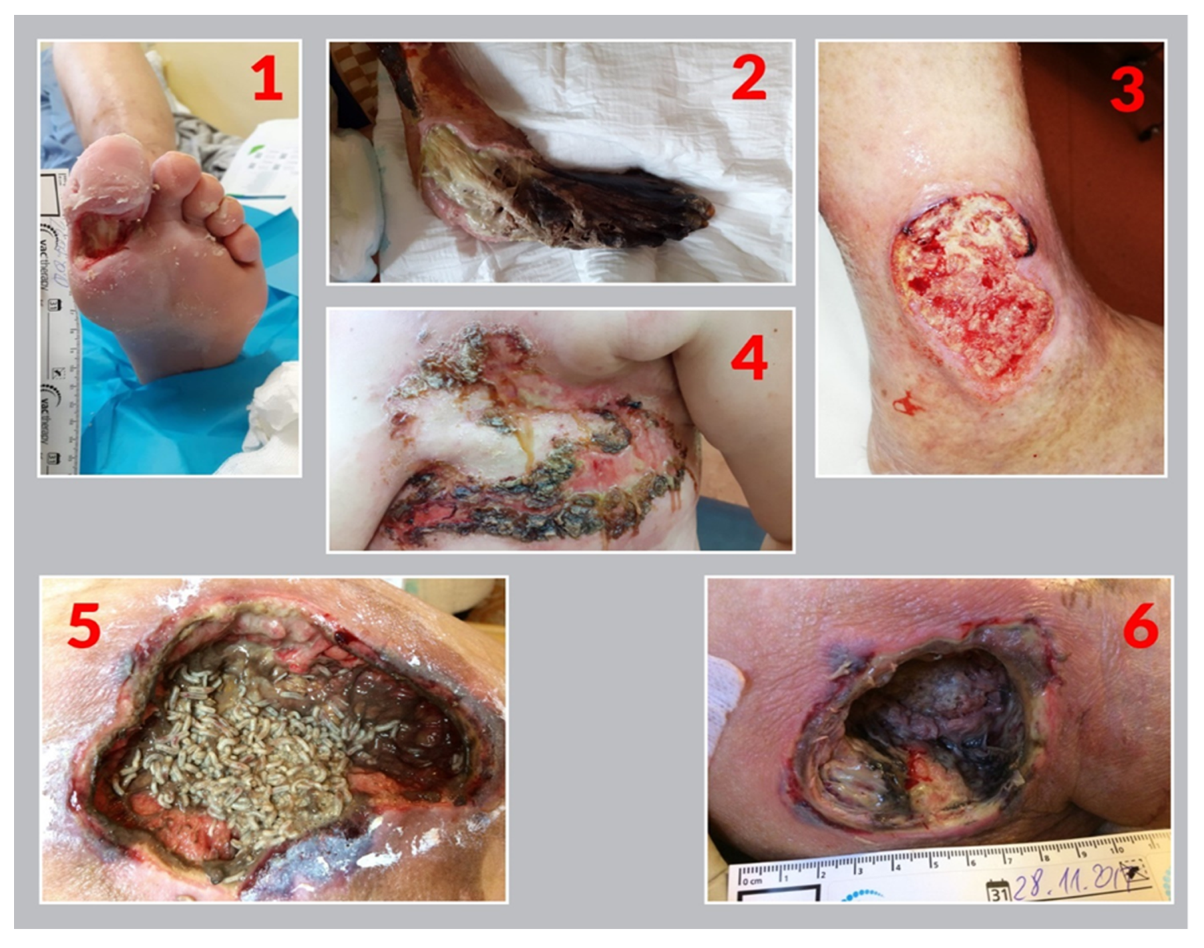
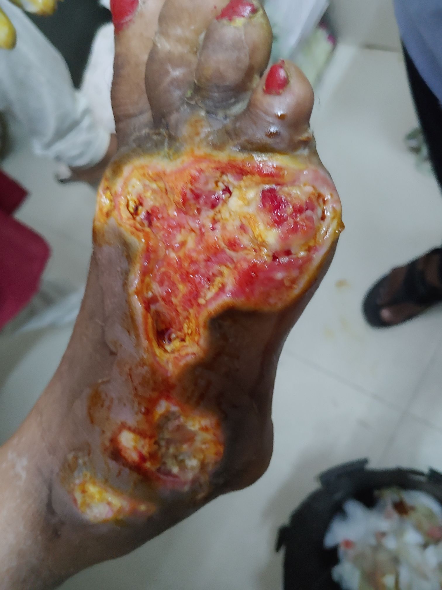
Use anatomical location-heel ankle sacrum coccyx etc. Diabetic foot ischemic with necrosis Figure 2. A controlled cohort study was done with 18 diabetic patients who had neuropathic non-healing leg wounds.
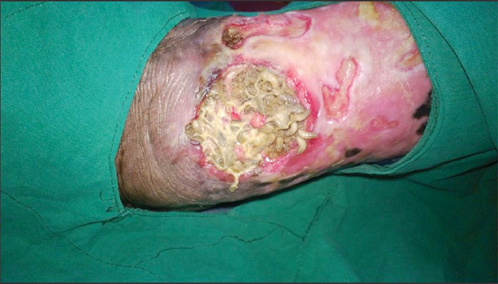
Diabetic foot ulcers are the consequence of multiple factors including peripheral neuropathy decreased blood supply high plantar pressures. Ischemic disease necrotic wound tissue Etiology. Necrotic diabetic foot Figure 3.
Risk factors include diabetes peripheral arterial disease. Compression bandages should be removed immediately if the person experiences a change in foot colour or temperature or increased pain. The maggots will not consume healthy tissue.
The larvae have a ferocious appetite for.

Infestation Of A Diabetic Foot By Wohlfahrtia Magnifica Sciencedirect
Ijerph Free Full Text Perception And Readiness To Undertake Maggot Debridement Therapy With The Use Of Lucilia Sericata Larvae In The Group Of Nurses Html

Resources Dr Biomaggot

Treatment Course Of Maggot Therapy A Before Maggot Therapy Necrotic Download Scientific Diagram
File Maggot Debridement Therapy On A Diabetic Foot Jpg Wikipedia
Medizzy Maggot Therapy

Diabetic Ulcer Maggots Removal Diabetic Foot Case Before After Youtube

Maggot Therapy An Overview Sciencedirect Topics

Reviving Hope By Using Of Maggot Debridement Therapy In Patients With Diabetic Foot Ulcer A Case Report Study Sciencedirect

Infected Diabetic Foot Wound Wagner S Grade 4 With Maggots Download Scientific Diagram
Medizzy Maggot Therapy
Freely Crawling Maggots Download Scientific Diagram

Use Of Lucilia Sericata Blowfly Maggots In The Treatment Of Diabetic Feet Threatened With Amputation Semantic Scholar
Diabetic Ulcer Maggots Removal Diabetic Foot Case Before After Youtube

Maggot Therapy An Alternative For Wound Infection The Lancet

A Retrospective Quality Improvement Review Of Maggot Debridement Therapy Outcomes In A Foot And Leg Ulcer Clinic Semantic Scholar
Using Maggots In Wound Care Part 2 Wound Care Advisor

Infected Charcot Foot Podiatrist India

David G Armstrong On Twitter Having A Wound Immediately Doubles One S Chances Of Dying At 10 Years Compared With Someone Without Diabetes Diabeticfoot Https T Co S9ntgputk2 Twitter


Comments
Post a Comment